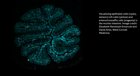
In the News

Two Weill Cornell Medicine Faculty Recognized with AAI Awards

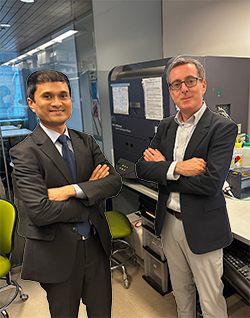
a composite image of tworesearchers

Two Weill Cornell Medicine faculty members, Dr. Gregory Sonnenberg and Dr. Melody Zeng, are recipients of prestigious awards from the American Association of Immunologists (AAI) for their accomplishments in the field of immunology.

Dr. Sonnenberg is the 2023 recipient of the AAI-BD Biosciences Investigator Award, recognizing his outstanding contributions to the field of immunology as a mid-career scientist, and Dr. Zeng is a recipient of an AAI ASPIRE Award, recognizing her work as an early-career immunologist and potential for advancing the field of immunology. The AAI has been dedicated to advancing immunology to improve health and fostering development opportunities for immunologists since it was founded in 1913.

Dr. Sonnenberg, the Henry R. Erle, MD-Roberts Family Associate Professor of Medicine is being honored for his laboratory’s research defining how the immune system controls health, immunity and inflammation in tissues lining various body surfaces, such as the gastrointestinal tract and respiratory system, and how immune system dysfunction can lead to gastrointestinal diseases, multiple sclerosis, asthma or cancer. For example, Dr. Sonnenberg and his lab discovered that immune cells called group 3 innate lymphoid cells (ILC3s) play a central role in regulating immunologic tolerance and healthy immune cell interactions with trillions of beneficial microbes in the gut, known as the microbiota. They also recently found that dysfunction of ILC3s may explain how colorectal cancer progresses and why this disease is resistant to checkpoint blockade immunotherapy.

“It is a tremendous honor to be chosen as the 2023 recipient of the AAI-BD Biosciences Investigator Award. I am also very humbled to be recognized by this important association and to be included among a list of prior recipients that are such phenomenal scientists,” said Dr. Sonnenberg, who is also a member of the Division of Gastroenterology & Hepatology and Jill Roberts Institute for Research in Inflammatory Bowel Disease at Weill Cornell Medicine. “This award also recognizes the significant contributions and dedication of talented individuals that I am lucky enough to have worked with in my lab over the years, as well as close collaborators and colleagues within our multidisciplinary network at Weill Cornell Medicine.”

Dr. Zeng, an assistant professor of immunology in pediatrics, is being recognized for her research and contribution to the growing collective evidence that the gut microbiota regulates the immune system. As a postdoctoral fellow at the University of Michigan School of Medicine, she was among the first to discover that a class of antibodies called IgG recognizes gut bacteria for maintenance of host-microbe symbiosis in the intestine. Dr. Zeng established her lab at Weill Cornell Medicine in 2019 and has continued to study the role of IgG in immune regulation by gut bacteria and its role in inflammatory and infectious disorders in early life. For example, her team recently discovered in a preclinical study that IgG antibodies transferred from mothers to infants through breast milk help shape infants’ gut bacteria and immunity against pathogenic bacteria that cause diarrheal illness.

“I am very honored and excited to be recognized along with the other recipients of this year’s AAI ASPIRE Award,” said Dr. Zeng, who is also a member of the Gale and Ira Drukier Institute for Children’s Health at Weill Cornell Medicine. “The annual AAI Meeting is a preeminent conference for immunologists, providing a platform for trainees to showcase their research and to network. It is also where I gave my first national meeting presentation as a student more than ten years ago, and it will be a sincere thrill to give a talk as an award recipient at this year’s event. This award also acknowledges the excellent training I received from my mentors, as well as the exciting research program that my amazing team works hard every day to build with tremendous support from many of my colleagues at Weill Cornell Medicine.”
The award ceremony will take place in May in Washington, D.C., at the organization’s 106th annual conference, where Drs. Sonnenberg and Zeng will present lectures about their research.

NIH Awards MERIT Grant for Inflammatory Bowel Disease Research

man in suit jacket sitting at lab hood

Dr. Gregory F. Sonnenberg, The Henry R. Erle, M.D.-Roberts Family Associate Professor of Medicine and head of basic research in Gastroenterology and Hepatology at Weill Cornell Medicine, has been awarded a $3.26 million, five-year MERIT grant from the National Institute of Allergy and Infectious Diseases (NIAID), part of the National Institutes of Health, to investigate the underlying mechanisms of inflammatory bowel disease (IBD).

The new grant, which has the possibility of extension for five additional years, will fund research on a novel pathway that protects the intestine from damage and inflammation driven by an immune-derived factor called tumor necrosis factor (TNF).

“It’s a huge honor to be considered and nominated for a NIH MERIT award, and such a privilege to be able to move this research forward with my laboratory over an extended period of time,” Dr. Sonnenberg said.

The MERIT program, a backronym for “Method for Extending Research In Time,” gives investigators with stellar records of research accomplishment a five-year award with the possibility of extending the initial award for up to five additional years without the need to undergo another competitive peer review.

Dr. Sonnenberg’s proposal was based on research his laboratory pioneered over the past decade characterizing key immune cell pathways in the intestine. It’s one of the major mysteries in the field. “TNF is normally beneficial, but what causes this shift for it to become a major mediator of chronic inflammatory diseases?” asked Dr. Sonnenberg, who is in the Division of Gastroenterology & Hepatology in the Weill Department of Medicine and a member of the Jill Roberts Institute for Research in Inflammatory Bowel Disease at Weill Cornell Medicine.

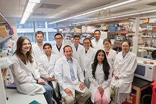
picture of Greg Sonnenberg and his lab members sitting in the lab

The Sonnenberg lab, with Dr. Greg Sonnenberg, seated second from right.


Many current therapies block TNF to reduce inflammation in chronic inflammatory diseases, including IBD, rheumatoid arthritis and psoriasis, but those treatments are only effective in a subset of patients, and even if successful, this therapy can lose efficacy over time. Dr. Sonnenberg’s team discovered that TNF-induced gut inflammation correlates with the depletion of group 3 innate lymphoid cells (ILC3s), a special class of cells abundant in the healthy intestine. “This provoked a hypothesis that these cells are keeping the TNF molecule in check and stopping it from driving tissue inflammation,” Dr. Sonnenberg said.

Experiments published last year in preclinical models confirmed that hypothesis and revealed that ILC3s are essential to protect the intestine from TNF-mediated damage and inflammation, which the team will explore further with support of this MERIT Award. Dr. Sonnenberg and his laboratory proposed a series of experiments to define the cellular and molecular signals by which ILC3s protect from TNF-induced inflammation. He also plans to investigate this pathway in both mouse models and patient-based organoids, complex human cell culture systems that model the guts of healthy individuals or patients with IBD.

Researchers cannot apply for MERIT grants directly. Dr. Sonnenberg and his laboratory originally applied for a standard “R01” grant from the NIH, which received a fundable score after peer-review and was subsequently nominated by NIH program staff for the MERIT award instead. Each year, NIAID issues about 15 new MERIT awards. The new award, which can double the length and amount of the grant, will take the investigation further, laying the groundwork for a much deeper understanding of IBD and paving the way for new therapeutic strategies.

Research reported in this newsroom story was supported by the National Institute of Allergy and Infectious Diseases of the National Institutes of Health under award number R37AI174468.

Gut Fungi's Lasting Impact on Severe COVID-19 Immune Response

closeup of monitor beside patient bed with patient on a respirator

Certain gut-dwelling fungi flourish in severe cases of COVID-19, amplifying the excessive inflammation that drives this disease while also causing long-lasting changes in the immune system, according to a new study led by investigators at Weill Cornell Medicine and NewYork-Presbyterian. This discovery identifies a group of patients who may benefit from specialized, but yet-to-be determined treatments.

Dr. Iliyan Iliev

Dr. Iliyan Iliev


Utilizing patient samples and preclinical models, the research team determined that the growth of fungi in the intestinal tract, particularly strains of Candida albicans yeast, trigger an upsurge in immune cells whose actions can exacerbate lung damage. Their findings, published in Nature Immunology on Oct. 23, also elucidate that patients retain a heightened immune response and immune memory against these fungi for up to a year after the resolution of SARS-CoV-2 infection. The research reveals a new dimension of the complex pathology unleashed by severe COVID-19, according to senior author Dr. Iliyan Iliev, an associate professor of immunology in medicine in the department of medicine, co-director of the Microbiome Core Lab and a member of the Jill Roberts Institute for Research in Inflammatory Bowel Disease at Weill Cornell Medicine. 

“Severe and long COVID-19 were not thought to involve fungal blooms in the intestines that, in addition to the virus, can impact patient’s immunity,” he said. 

Dr. Iliev, an immunologist who studies the microbiome and the chronic inflammatory conditions targeting the gastrointestinal tract, pivoted to COVID-19 during the pandemic. As researchers gained a better handle on the new viral infections, it became clear that, in COVID-19 as in inflammatory bowel disease, the body’s own inflammatory immune response causes harm.

Dr. Takato Kusakabe

Dr. Takato Kusakabe


To investigate this errant immune response, Dr. Iliev and Dr. Takato Kusakabe, a postdoctoral fellow and a first author in the study, worked with numerous colleagues to acquire three large clinical cohorts of COVID-19 patients and develop a mouse model to study the disease. They collaborated with members of the Weill Department of Medicine and the Department of Pathology and Laboratory Medicine at Weill Cornell Medicine, including Dr. Stephen JosefowitzDr. Mirella SalvatoreDr. Melissa Cushing, Dr. Lars Westblade, and Dr. Adolfo García-Sastre, a professor of microbiology and director of the Global Health and Emerging Pathogens Institute of the Icahn School of Medicine at Mount Sinai.

How Gut Fungi Harm the Lungs

The team first made the connection when analysis of blood samples from patients at New York-Presbyterian/Weill Cornell Medical Center diagnosed with severe COVID-19 unveiled the presence of antibodies tuned to attack fungi common to the gut. The researchers then found that populations of yeast, and one species in particular, Candida albicans, increased in the intestines of the patients during the course of severe COVID-19. 

When they looked at the patients’ immune systems, the researchers found a parallel increase in immune cells called neutrophils. In severe COVID-19, excessive numbers of neutrophils appear in the lungs, where their activity worsens the inflammatory response already damaging these organs. 

Turning to preclinical models, the investigators found that mice bearing fungi from patients with severe COVID-19 produced more neutrophils in their blood and lungs, and had signs of heightened inflammation when infected with SARS-CoV-2. However, giving them an antifungal drug reduced these effects. 

The Immune System Remembers

From within patients’ blood samples, researchers also uncovered evidence of persistent changes to the immune system they believe are related to a condition known as long COVID-19, in which symptoms linger, or new ones develop, after an infection has cleared.  

When the team examined patients’ blood up to a year afterward, they found it still contained anti-fungal antibodies. In addition, when they looked at the stem cells that give rise to neutrophils, the researchers found that these progenitors are primed to respond to fungi. They found that an immune protein called IL-6 that these fungi induce, appears to bolster both the neutrophils and the antibodies.   

Further experiments showed that blocking IL-6 in the patients or in mice dampened this immunological memory, causing the presence of neutrophils and antibodies to wane. 

While these results do not have immediate implications for treating severe or long COVID-19, they suggest new opportunities to tailor therapy, according to Dr. Iliev. For example, anti-fungal antibodies could potentially serve as a marker to identify patients who might benefit from a therapy that targets the fungi or the immunological changes they instigate. Or, assuming further research supports it, the antibodies’ presence could indicate someone might be at risk for long COVID-19. The team’s discoveries may also have relevance beyond COVID-19, said Dr. Iliev, who notes this research could open new avenues of exploration for the treatment of other infectious and inflammatory diseases.

Many Weill Cornell Medicine physicians and scientists maintain relationships and collaborate with external organizations to foster scientific innovation and provide expert guidance. The institution makes these disclosures public to ensure transparency. For this information, see the profile for Dr. Melissa Cushing.

Research reported in this newsroom story was supported by the National Institutes of Health grants P30CA016087, R01AI143861, R01AI143861-02S1, R01AI160706, R01DK130425, U19AI135972, U19AI168631, U19AI142733, DK113136, DK121977 and AI137157. Additional support was provided by the Jill Roberts Institute for Research in Inflammatory Bowel Disease, the Leona M. and Harry B. Helmsley Charitable Trust, the Irma T. Hirschl Career Scientist Award, Crohn’s and Colitis Foundation, and the Burrough Welcome Trust PATH Award. Dr. Iliev is a fellow of the CIFAR program Fungal Kingdom: Threats and Opportunities.

New Consortium Aims to Transform Understanding of How the Human Body Senses Health and Disease


a yellow scientific image against a black backdrop

New York, NY (November 9, 2023) - The Allen Discovery Center (ADC) for Neuroimmune Interactions at the Icahn School of Medicine at Mount Sinai hopes to revolutionize the field of medicine with a groundbreaking project to understand how the human body senses health and disease.

Spearheaded by Dr. Brian S. Kim of Mount Sinai and co-led by Dr. David Artis of Weill Cornell Medicine, this new multidisciplinary research center brings together leading experts studying the intersection of immunology and neuroscience. This pioneering effort aims to reshape our understanding of fundamental biology and human disease, with the near-term goal of changing the way we treat chronic diseases.

Headshot of a man sitting in a green chair

Dr. David Artis. Credit: John Abbott


The Allen Discover Center for Neuroimmune Interactions is funded at $10 million over four years by the Paul G. Allen Family Foundation, with a total potential for $20 million over eight years. The Allen Discovery Center program is advised by The Paul G. Allen Frontiers Group.

The motivation behind this center is rooted in the profound advances achieved in immunology and neuroscience over the past decade. These breakthroughs have unveiled the intricate communication between the immune and nervous systems. The team’s mission is to explore the role of immune cells as environmental and internal sensors influencing various aspects of human behavior and to recognize the sensory nervous system's newfound capacity to act as an immune organ.

“Together, our combined expertise aims to advance fundamental neuroimmunology, with a commitment to translating our findings into revolutionary therapies rapidly,” said Dr. Kim. “For example, many diseases arise from the immune system going awry. However, we are now finding that the nervous system communicates closely with the immune system to sense inflammation in ways we never imagined. We are also seeing how different organs communicate through this unique network. We can envision a future in which we treat psoriasis by tuning nerves in the skin, anxiety by modulating gut inflammation, and improving cancer therapy by limiting bad nerves from sprouting into the tumor.”

Physician-scientist in the lab

Dr. Brian Kim. Credit: Mount Sinai Health Syst


Dr. Kim, vice chair of research and professor of dermatology, Icahn Mount Sinai, and director of the Mark Lebwohl Center for Neuroinflammation and Sensation at Mount Sinai, pioneered the understanding of peripheral neurons as sensors of inflammation leading to FDA-approved itch therapies. He will lead this collaboration, joined by an exceptional team of investigators from Icahn Mount Sinai, Weill Cornell Medicine, NYU Langone Health, and Yale School of Medicine.

“Collaborating with The Paul G. Allen Frontiers Group to launch the Allen Discovery Center for Neuroimmune Interactions here in New York City allows us to bring together outstanding investigators from Icahn Mount Sinai, NYU Langone Health, Weill Cornell Medicine, and Yale School of Medicine, providing a tremendous opportunity to leverage cutting-edge technologies to provide new insights into how the nervous and immune systems communicate with each other to regulate immunity, inflammation, and tissue homeostasis,” said Dr. Artis, who is director of the Jill Roberts Institute for Research in Inflammatory Bowel Disease and director of the Friedman Center for Nutrition and Inflammation at Weill Cornell Medicine. “The outcome of these studies will have implications for our understanding of the pathophysiology of a number of diseases, from asthma and allergy to autoimmunity and cancer.”
About the Mount Sinai Health System

Mount Sinai Health System is one of the largest academic medical systems in the New York metro area, with more than 43,000 employees working across eight hospitals, more than 400 outpatient practices, more than 300 labs, a school of nursing, and a leading school of medicine and graduate education. Mount Sinai advances health for all people, everywhere, by taking on the most complex health care challenges of our time—discovering and applying new scientific learning and knowledge; developing safer, more effective treatments; educating the next generation of medical leaders and innovators; and supporting local communities by delivering high-quality care to all who need it. Through the integration of its hospitals, labs, and schools, Mount Sinai offers comprehensive health care solutions from birth through geriatrics, leveraging innovative approaches such as artificial intelligence and informatics while keeping patients’ medical and emotional needs at the center of all treatment. The Health System includes approximately 7,400 primary and specialty care physicians; 13 joint-venture outpatient surgery centers throughout the five boroughs of New York City, Westchester, Long Island, and Florida; and more than 30 affiliated community health centers. Hospitals within the System are consistently ranked by Newsweek’s® “The World’s Best Smart Hospitals, Best in State Hospitals, World Best Hospitals and Best Specialty Hospitals” and by U.S. News & World Report's® “Best Hospitals” and “Best Children’s Hospitals.” The Mount Sinai Hospital is on the U.S. News & World Report® “Best Hospitals” Honor Roll for 2023-2024.

About The Paul G. Allen Frontiers Group

The Paul G. Allen Frontiers Group, a division of the Allen Institute, is dedicated to exploring the landscape of bioscience to identify and foster ideas that will change the world. The Frontiers Group recommends funding to The Paul G. Allen Family Foundation, which then invests through award mechanisms to accelerate our understanding of biology, including: Allen Discovery Centers at partner institutions for leadership-driven, compass-guided research; and Allen Distinguished Investigators for frontier explorations with exceptional creativity and potential impact. The Paul G. Allen Frontiers Group was founded in 2016 by the late philanthropist and visionary Paul G. Allen. For more information, visit alleninstitute.org/division/frontiers-group/

About Weill Cornell Medicine

Weill Cornell Medicine is committed to excellence in patient care, scientific discovery and the education of future physicians in New York City and around the world. The doctors and scientists of Weill Cornell Medicine—faculty from Weill Cornell Medical College, Weill Cornell Graduate School of Medical Sciences, and Weill Cornell Physician Organization—are engaged in world-class clinical care and cutting-edge research that connect patients to the latest treatment innovations and prevention strategies. Located in the heart of the Upper East Side’s scientific corridor, Weill Cornell Medicine’s powerful network of collaborators extends to its parent university Cornell University; to Qatar, where Weill Cornell Medicine-Qatar offers a Cornell University medical degree; and to programs in Tanzania, Haiti, Brazil, Austria and Turkey. Weill Cornell Medicine faculty provide exemplary patient care at NewYork-Presbyterian/Weill Cornell Medical Center, NewYork-Presbyterian Westchester Behavioral Health Center, NewYork-Presbyterian Lower Manhattan Hospital, NewYork-Presbyterian Queens and NewYork-Presbyterian Brooklyn Methodist Hospital. Weill Cornell Medicine is also affiliated with Houston Methodist. For more information, visit weill.cornell.edu.

Pain-Sensing Gut Neurons Protect Against Inflammation

Neurons that sense pain protect the gut from inflammation and associated tissue damage by regulating the microbial community living in the intestines, according to a study from researchers at Weill Cornell Medicine.

The researchers, whose report appears Oct. 14 in Cell, found in a preclinical model that pain-sensing neurons in the gut secrete a molecule called substance P, which appears to protect against gut inflammation and related tissue damage by boosting the population of beneficial microbes in the gut. The researchers also found that these pain-sensing nerves are diminished in number, with significant disruptions to their pain-signaling genes, in people who have inflammatory bowel disease (IBD).

“These findings reshape our thinking about chronic inflammatory disease, and open up a whole new approach to therapeutic intervention,” said study senior author Dr. David Artis, director of the Jill Roberts Institute for Research in Inflammatory Bowel Disease, director of the Friedman Center for Nutrition and Inflammation and the Michael Kors Professor of Immunology at Weill Cornell Medicine.

a man posing for a picture

Dr. David Artis


The study’s first author, Dr. Wen Zhang, a postdoctoral researcher in the Artis laboratory, added, “Defining a previously unknown sensory function for these specific neurons in influencing the microbiota adds a new level of understanding to host-microbiota interactions.”

IBD covers two distinct disorders, Crohn’s disease and ulcerative colitis, and is believed to affect several million people in the United States. Typically it is treated with drugs that directly target elements of the immune system. Scientists now appreciate that gut-dwelling bacteria and other microbes also help regulate gut inflammation.

As Dr. Artis’s laboratory and others have shown in recent years, the nervous system, which is “wired” into most organs, appears to be yet another powerful regulator of the immune system at the body’s barrier surfaces. In the new study, Dr. Artis and his team specifically examined pain neurons that innervate—extend their nerve endings into—the gut.

a woman smiling for a portrait

Dr. Wen Zhang


These gut-innervating pain neurons, whose cell bodies sit in the lower spine, express a surface protein called TRPV1, which serves as a receptor for pain-related signals. TRPV1 can be activated by high heat, acid, and the chili-pepper compound capsaicin, for example—and the brain translates this activation into a sense of burning pain. The researchers found that silencing these TRPV1 receptors in gut nerves, or deleting TRPV1-expressing neurons, led to much worse inflammation and tissue damage in IBD mouse models, whereas activating the receptors had a protective effect.

The investigators observed that the worsened inflammation and tissue damage in TRPV1-blocked mice were associated with changes in the relative populations of different species of gut bacteria. When this altered bacterial population was transplanted into normal mice, it caused the same worsened susceptibility to inflammation and damage. By contrast, broad-spectrum antibiotic treatment could reverse this susceptibility even in TRPV1-blocked mice. This result demonstrated that TRPV1-expressing nerves protect the gut mainly by helping to maintain a healthy gut microbe population. To continue reading, click here.

Fungal Association with Tumors May Predict Worse Outcomes

The presence of some fungal species in tumors predicts—and may even help drive—worse cancer outcomes, according to a study from Weill Cornell Medicine and Duke University researchers.

The study, which appears Sept. 29 in Cell, provides a scientific framework to develop tests that delineate specific fungal species in tumors that are relevant for prediction of cancer progression and therapy. The results also point to the possibility of using antifungal treatments to augment conventional cancer treatments in some cases.

“These findings open up a lot of exciting research directions, from the development of diagnostics and treatments to studies of the detailed biological mechanisms of fungal relationships to cancers,” said senior author Dr. Iliyan Iliev, associate professor of immunology in medicine in the Division of Gastroenterology and Hepatology and a member of the Jill Roberts Institute for Research in Inflammatory Bowel Disease at Weill Cornell Medicine.

a man posing for a picture

Dr. Iliyan Iliev. Credit: Jennifer Conrad


The first author of the study was Anders Dohlman, a doctoral student in biomedical engineering at Duke University.

The idea that viruses and bacteria can trigger or accelerate cancer development is now well established. However, little is known about the cancer-related roles of fungi—which, like bacteria and viruses, colonize the gut, lungs, skin and other barrier tissues, interact with the immune system, and sometimes cause disease.

In the new study, researchers catalogued fungal species and their associations with different cancers by analyzing The Cancer Genome Atlas, the largest well-annotated genomic database of human tumors.

The analysis revealed that the DNA of certain fungal species are relatively abundant in some tumor types. These species include, in gastrointestinal tumors, Candida tropicalis and Candida albicans, which causes thrush and yeast infections; in lung tumors, species of the fungal genus Blastomyces; and in breast tumors, species of the fungus Malassezia.

The researchers devised sophisticated computational methods to exclude fungal DNA likely to have originated from laboratory contamination, and were able to confirm, in particular, the presence of live Candida species in colorectal tumor samples.

Their analysis linked higher levels of Candida in gastrointestinal tumors to tumor gene activity promoting inflammation and reduced cell-to-cell adhesion—features associated with cancer’s late-stage spread to distant organs, known as metastasis. Higher Candida levels for such tumors were also directly associated with a greater rate of metastasis.

The findings, according to the researchers, suggest that high levels of particular fungi in tumor biopsies might someday be used as biomarkers, indicating, for example, a higher metastasis risk—which in turn could lead to the choice of more effective treatment. To continue reading, click here.

Discovery Reveals How the Immune System Tolerates Friendly Gut Bacteria

Immune cells called group 3 innate lymphoid cells (ILC3s) play an essential role in establishing tolerance to symbiotic microbes that dwell in the human gastrointestinal tract, according to a study led by researchers at Weill Cornell Medicine.

The discovery, reported Sept. 7 in Nature, illuminates an important aspect of gut health and mucosal immunity—one that may hold the key to better treatments for inflammatory bowel disease (IBD), colon cancer and other chronic disorders.

“As part of this study, we define a novel pathway that drives immune tolerance to microbiota in the gastrointestinal tract,” said senior author Dr. Gregory F. Sonnenberg, associate professor of microbiology and immunology in medicine and head of basic research in the Division of Gastroenterology & Hepatology, and a member of the Jill Roberts Institute for Research in Inflammatory Bowel Disease at Weill Cornell Medicine. “This is a fundamental advance in our understanding of mucosal immunity and may hold the key to understanding what goes wrong when the immune system begins to inappropriately attack microbiota in diseases such as IBD.”

two men posing for a photo

Drs. Mengze Lyu and Gregory Sonnenberg


Scientists have long known that trillions of bacteria, fungi, and other microbes dwell symbiotically in the intestines of mammals. The mechanism by which the immune system normally tolerates these “beneficial” gut microbes, instead of attacking them, has not been well understood. But there is evidence that this tolerance breaks down in IBD, leading to harmful flareups of gut inflammation. Thus, a detailed understanding of gut immune tolerance could enable the development of powerful new treatments for IBD—a class of diseases that include Crohn’s disease and ulcerative colitis, which affect several million individuals in the United States alone.

In the study, Dr. Sonnenberg and colleagues, including lead author Dr. Mengze Lyu, a postdoctoral researcher in the Sonnenberg lab, used single-cell sequencing and fluorescent imaging techniques to delineate immune cells in the mesenteric lymph nodes that drain the intestines of healthy mice. They focused on cells expressing a transcription factor, RORγt, which are known to drive either inflammation or tolerance in response to microbes that colonize the intestine. The dominant immune cell types in these tissues, they found, were T cells and ILC3s. The latter are a family of immune cells that represent an innate counterpart of T cells, and work as a first line of defense in mucosal tissues such as the intestines and lungs.

In close collaboration with researchers at the University of Birmingham, UK, the scientists observed that in lymph node regions called interfollicular zones, ILC3s are in close association with a specific type of T cell, called RORγt+ regulatory T cells (Tregs), which are adapted to dial down inflammation and immune activity to promote tolerance in the gut. To continue reading, please click here.

Scientists Identify a Key Molecular Protector of Gut Health

A protein called Zbtb46, expressed by specialized immune cells, has a major role in protecting the gastrointestinal tract from excessive inflammation, according to a study from researchers at Weill Cornell Medicine.
The finding, which appears July 13 in Nature, is a significant advance in the understanding of how the gut maintains health and regulates inflammation, which could lead to better strategies for treating diseases like inflammatory bowel disease (IBD).

“We’ve known that there are related families of immune cells in the gut that can either protect from inflammation or at other times be major drivers of inflammation,” said senior author Dr. Gregory Sonnenberg, associate professor of microbiology and immunology in medicine in the Division of Gastroenterology & Hepatology, and a member of the Jill Roberts Institute for Research in Inflammatory Bowel Disease at Weill Cornell Medicine. “This new finding helps us understand how these cells are regulated to optimally promote intestinal health and prevent inflammation.”
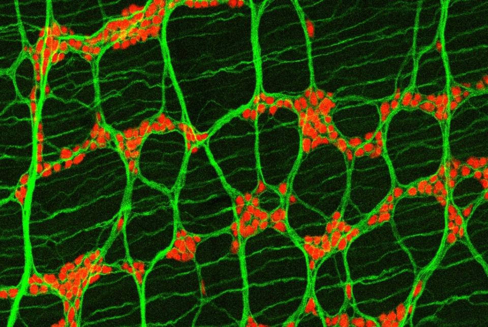
IBD, which includes Crohn’s disease and ulcerative colitis, affects several million people in the United States. These chronic inflammatory disorders target the gut, can be seriously debilitating, and treatments may not work well for some patients—mainly because scientists don’t have a complete picture of what is driving these diseases and how the sophisticated immune cell networks in the gut support tissue health.

Dr. Sonnenberg’s laboratory has been advancing the science of gut immunity with studies of recently identified immune cells called ILC3s. These “innate lymphoid cells” are related to T cells and B cells, and clearly have important roles in protecting the gut and other organs from excess inflammation. However, in the context of IBD or colorectal cancer they become altered. In general, scientists have wanted to know more about how these cells work.

a man and woman smiling for a portrait

Drs. Gregory Sonnenberg and Wenqing Zhou. Provided by Sonnenberg lab


To this end, Dr. Sonnenberg and his team, including first author Dr. Wenqing Zhou, a postdoctoral researcher in the Sonnenberg Laboratory, set out to make a detailed catalog of ILC3s and other related immune cells residing in the large intestine of mice, using relatively new single-cell sequencing techniques.

A surprise finding was that a subset of ILC3s express Zbtb46, an anti-inflammatory protein that prior studies had suggested is produced only in dendritic cells, a very different type of immune cell. The researchers showed in experiments with mice that ILC3s expressing Zbtb46 have a strong ability to restrain inflammation following gut infection. When they blocked Zbtb46 expression in ILC3s, gut infection led to signs of severe inflammation, including a rise in the numbers of other gut immune cells promoting inflammation. To read further, please click here

Toxin-producing Yeast Strains in Gut Fuel IBD

Individual Candida albicans yeast strains in the human gut are as different from each other as the humans that carry them, and some C. albicans strains may damage the gut of patients with inflammatory bowel disease (IBD), according to a new study from researchers at Weill Cornell Medicine. The findings suggest a possible way to tailor treatments to individual patients in the future.

The researchers, who report their findings March 16 in Nature, used an array of techniques to study strains, or genetic variants, of Candida from the colons of people with or without ulcerative colitis, a chronic, relapsing and remitting inflammatory disorder of the colon and rectum and one of the main forms of IBD. They found that certain strains, which they call “high-damaging,” produce a potent toxin called candidalysin that damages immune cells.

a man posing for a picture

Dr. Iliyan Iliev


“Such strains retained their “high-damaging” properties when they were removed from the patient’s gut and triggered pro-inflammatory immunity when colonized in mice, replicating certain disease hallmarks,” said senior author Dr. Iliyan Iliev, an associate professor of immunology in medicine in the Division of Gastroenterology and Hepatology and a scientist in the Jill Roberts Institute for Research in Inflammatory Bowel Disease at Weill Cornell Medicine.

IBD affects approximately 3.1 million people in the United States and can greatly impair patients’ quality of life. Such patients rely on a handful of available therapies, but treatments may not always be effective. The new study has suggested one reason steroids, a commonly used treatment, may not work; treating mice with the drug to suppress intestinal inflammation failed in the presence of “high-damaging” C. albicans strains.

“Our findings suggest that C. albicans strains do not cause spontaneous intestinal inflammation in a host with intact immunity,” Dr. Iliev said. “But they do expand in the intestines when inflammation is present and can be a factor that influences response to therapy in our models and perhaps in patients.”

Most studies of the human microbiome in healthy individuals and those with IBD have focused on bacteria and viruses, but recent research by Dr. Iliev and others has illuminated the contributions of fungi to the effects of microbes on humans and mice. They have found that intestinal fungi play an important role in regulating immunity at surfaces exposed to the outside, such as the intestines and lungs, due to their potent immune-stimulating characteristics. While the collective community of fungi in the body, known as the mycobiota, has been linked to several diseases, including IBD, researchers previously had not understood the mechanisms by which the mycobiota contribute to inflammation in the gut. To continue reading article, click here.

A New and Versatile Genetic Manipulation Pipeline for Studying Nonmodel Gut Bacteria

Scientists at the Jill Roberts Institute for Research in Inflammatory Bowel Disease at Weill Cornell Medicine have developed a pipeline that enables genetic manipulation of nonmodel gut bacteria. The pipeline will allow scientists to study the biological roles of these bacteria, which are increasingly recognized as key factors in health and disease, at the single-gene level.

Scientists have developed advanced genetic tools for some model gut bacteria, such as E. coli, but have lacked the necessary tools for a large group of gut bacteria called Firmicutes/Clostridia that are dominant in a healthy human gut. In the study, published Jan. 19 in Cell, the researchers have developed gene-modification techniques for multiple nonmodel gut bacteria, from more than five different phyla including Firmicutes/Clostridia.

The researchers demonstrated the potential of their new genetic tools by using them to study the role of a key gut bacterial gene in regulating colon inflammation.

"Gut bacteria play a major role in the onset and development of human diseases like inflammatory bowel disease and cancers," said Dr. Chun-Jun Guo, assistant professor of immunology in medicine in the Department of Gastroenterology and Hepatology and a scientist in the Jill Roberts Institute for Research in Inflammatory Bowel Disease at Weill Cornell Medicine. “We were able not only to develop genetic manipulation tools for previously 'intractable' gut bacteria, but also to discover an interesting role for a microbial gene in the host using these tools.”

Gut bacteria have generally evolved symbiotic relationships, also known as commensal, with their animal hosts, including humans. Although scientists over the last two decades have been increasingly aware of gut microbes' importance in immune function and overall health—and in promoting disease when they are disrupted, for example by antibiotic use or poor diets—the development of genetic tools for manipulating these microbes has not kept pace. To read further, click here.

Two Weill Cornell Medicine Faculty Members Inducted into ASCI

Two Weill Cornell Medicine physician-scientists, Dr. Randy Longman and Dr. Robert Schwartz, have been elected as members of the American Society for Clinical Investigation.

The American Society for Clinical Investigation (ASCI) is one of the nation’s oldest nonprofit medical honor societies and focuses on the unique role of physician-scientists in research, clinical care and medical education. It is comprised of more than 3,000 physician-scientists serving in the upper ranks of academic medicine and industry. Members are leaders in their fields in translating innovative laboratory findings into clinical advancements. Drs. Longman and Schwartz join 93 other new members elected this year and will be officially inducted at the organization’s annual meeting in April.

a man in a suit posing for a picture

Dr. Randy Longman


Dr. Randy Longman is an associate professor of medicine in the Division of Gastroenterology and Hepatology at Weill Cornell Medicine and director of the Jill Roberts Center for Inflammatory Bowel Disease at Weill Cornell Medicine and NewYork-Presbyterian/Weill Cornell Medical Center. His research focuses on interactions between gut microbes and the immune system in inflammatory bowel disease (IBD). His work has identified specific strains of bacteria that are involved and how they relay signals to the immune system to activate inflammation in patients with Crohn’s disease and ulcerative colitis, the most common forms of IBD.

Dr. Longman is currently leading a clinical trial evaluating whether dietary fiber supplementation may improve the efficacy of fecal microbiota transplantation (FMT) in patients with moderate ulcerative colitis. Supported by the National Institute of Diabetes and Digestive and Kidney Diseases, the trial is based on his earlier work that identified specific microbes associated with clinical responses to FMT. Dr. Longman’s research is funded by several organizations, including the National Institutes of Health and the Crohn’s and Colitis Foundation.

“It’s very meaningful to be elected as a member of the ASCI, knowing my peers recognize my work has contributed toward moving the needle forward,” said Dr. Longman, who is also a member of the Jill Roberts Institute for Research in Inflammatory Bowel Disease. “As a physician-scientist, the most important thing for me is that my research discoveries may translate into clinical treatments that help patients.” To read further, click here.

Preclinical Study Finds Gut Fungi Influence Neuroimmunity and Behavior

A specific group of fungi residing in the intestines can protect against intestinal injury and influence social behavior, according to new preclinical research by scientists at Weill Cornell Medicine. The findings extend a growing body of work identifying a "gut-immunity-brain axis," a signaling system that may have a wide range of effects on physiology in both health and disease, influenced not only by the body's own cells but also the resident microbes.

The study, published Feb. 16 in Cell, reveals a novel set of molecular signals connecting fungi in the gut to their host’s cells throughout the body, including immune cells and neurons.

“We have made a direct link between a major immune pathway induced by fungi in the lining of the intestine and signals in the nervous system that impact animal behavior,” said senior author Dr. Iliyan Iliev, associate professor of immunology in medicine in the Division of Gastroenterology and Hepatology and a member of the Jill Roberts Institute for Research in Inflammatory Bowel Disease at Weill Cornell Medicine.
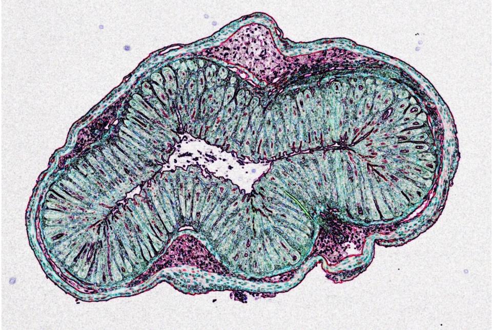
Drs. Dilek ColakMelanie Johncilla and Megan Allen from Weill Cornell Medicine and Dr. Rhonda K. Yantiss from Weill Cornell Medicine and NewYork-Presbyterian also contributed to this study.

The lining of the intestine must balance conflicting needs, absorbing water and nutrients from food while acting as a barrier to prevent the vast population of microbes in the gut from invading the bloodstream. Examining this system in a mouse model, the scientists mapped the locations of different fungi within the intestine and found that a unique consortium of fungi tends to accumulate at specific sites near the gut epithelium, or lining, suggesting that these species have colonized the gut and interact closely with the nearby epithelial cells.

image of two people in front of flow cytometry

Dr. Irina Leonardi (left) and Dr. Iliyan Iliev. Photo credit: Jennifer Conrad.


Mice carrying some of these fungi enjoyed better protection against events that can disrupt the intestinal barrier, such as intestinal injury and bacterial infection. "There was fortification of those barrier functions when we added that specific fungal community to mice," Dr. Iliev said.

Improving intestinal barrier integrity wasn't the only effect of the fungi. In separate experiments, the team found that mice carrying the fungal community in their gut displayed more social behavior than animals without these fungi. To read further, click here.

Study Offers Insight into Immune Mechanisms of Inflammatory Disease

ILC2 and goblet cells in intestines

Innate lymphoid cells are a recently discovered family of white blood cells that reside in the skin, gastrointestinal tract, airways and other barrier tissues of the body. Group 2 innate lymphoid cells (ILC2s) have an essential role in protecting these tissues from parasitic infections as well as damage associated with allergic inflammation and asthma, according to a new study led by Weill Cornell Medicine researchers.

The finding resolves a controversy about the possible redundancy of ILC2s with other cells in the body. The study also suggests that a unique set of regulatory networks controlled by neurons in the gut may be viable targets for future drug therapies to combat chronic inflammatory diseases including asthma, allergy and inflammatory bowel disease (IBD).

The study, published Nov. 2 in Nature, shows that although ILC2s have many functional similarities to immune cells called T helper type 2 cells (Th2 cells), the latter cell type cannot adequately compensate for loss of the protective response of ILC2s against parasitic worm infection in the gut as well as gut inflammation. Underscoring the clinical relevance of the study, the researchers found evidence that ILC2s in humans respond in a manner similar to mouse ILC2s.

a man posing for a picture

Dr. David Artis


“This advances our understanding of the complexity of the immune system, and gives us a potential new set of targets for future therapies,” said study senior author Dr. David Artis, director of the Jill Roberts Institute for Research in Inflammatory Bowel Disease, director of the Friedman Center for Nutrition and Inflammation and the Michael Kors Professor of Immunology at Weill Cornell Medicine.

ILC2s are part of a family of cells, innate lymphoid cells, that were discovered by multiple groups only about 12 years ago. With their strong presence in barrier tissues, innate lymphoid cells are generally considered to serve as sentinels and first responders against various types of infection. But scientists also recognize that ILCs may hold the keys to understanding common inflammatory and autoimmune conditions such as asthma and IBD.

It is thought that both ILC2s and Th2 cells evolved at least in part to defend the body from parasitic worm infections, biting insects and other environmental triggers. When triggered by such challenges, both help marshal what is called a type 2 immune response. These similarities have led researchers to suggest that they are functionally almost the same but ILC2s specialize in earlier, more localized responses, whereas T cells are more blood-borne and mobile, concentrating in multiple tissues where needed. However, in the new study, the researchers found that ILC2s have an essential immune role rather than being redundant as type 2 immune responders. 

When ILC2s and Th2 cells are activated by a worm infection, they both produce an anti-worm, tissue-protecting protein called amphiregulin (AREG). To determine if Th2 cells can compensate for loss of this protein from ILC2s, the researchers engineered mice in which AREG production is selectively deleted in ILC2s, but not in Th2 cells. They found that these mice were more susceptible to parasitic worm infection in the gut due to reduced capacity to mount an anti-parasitic immune response, compared with mice with normal ILC2s. The mice lacking ILC2 AREG were also much more susceptible to gut damage from inflammation.

Dr. Hiroshi Yano

Dr. Hiroshi Yano


“This finding clarifies that ILC2s are playing the major role in this tissue protective response—without them the response is inadequate,” said study co-first author Dr. Hiroshi Yano, a postdoctoral research associate in the Artis laboratory.

Clarifying the functional importance of a major immune cell type is a significant achievement in basic immunology and the results of the study also suggest clinical applications. The researchers showed that the ILC2 immune response, either to worm infection or inflammatory gut damage, is selectively controlled by a signaling molecule produced by neurons in the gut. Giving the molecule to mice with experimental gut inflammation boosted AREG production in ILC2s and protected the animals from gut damage. Preliminary experiments with gut ILC2s taken from patients with inflammatory bowel disease showed that the molecule could boost the protective response in the human cells as well. These findings suggest that neurons in the gut communicate with ILC2s to generate a protective response that cannot be replaced by other immune cells, thus offering new therapeutic opportunities, Dr. Artis said.

This work was supported in part by the National Institutes of Health (DK126871, AI151599, AI095466, AI095608, AI142213, AR070116, AI172027, DK132244), a WCM Department of Pediatrics Junior Faculty Pilot Award, the Jill Roberts Center Pilot Award for Research in IBD, a Thomas C. King Pulmonary Fellowship, the LEO foundation, Cure for IBD, Jill Roberts Institute, the Sanders Family, and the Rosanne H. Silbermann Foundation. 

A Common Dietary Fiber Promotes Allergy-Like Immune Responses in Preclinical Studies

goblet cells

A type of dietary fiber called inulin, commonly used in health supplements and known to have certain anti-inflammatory properties, can also promote an allergy-related type of inflammation in the lung and gut, and other parts of the body, according to a preclinical study from researchers in the Friedman Center for Nutrition and Inflammation and Jill Roberts Institute for Inflammatory Bowel Disease at Weill Cornell Medicine and in the Boyce Thompson Institute on Cornell’s Ithaca campus.

The study, published Nov. 2 in Nature, found that dietary inulin fiber alters the metabolism of certain gut bacteria, which in turn triggers what scientists call type 2 inflammation in the gut and lungs. This type of inflammation is thought to have evolved in mammals chiefly to defend against parasitic worm (“helminth”) infections, and is also part of normal wound-healing, although its inappropriate activation underlies allergies, asthma and other inflammatory diseases.

 John Abbott

Dr. David Artis photo. Credit: John Abbott


“There’s a lot to think about here, but, in general, these findings broaden our understanding of the relationship between diet, immunity, and the normally beneficial microorganisms that constitute our microbiota and colonize our bodies,” said study co-senior author Dr. David Artis, director of the Friedman Center for Nutrition and Inflammation and the Michael Kors Professor of Immunology at Weill Cornell Medicine.
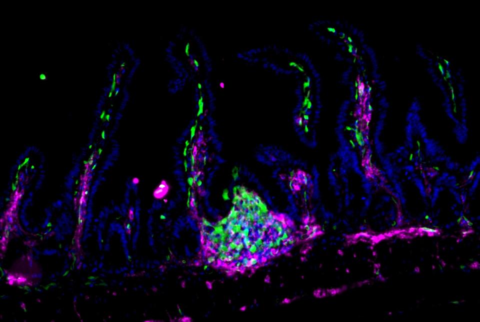
a man posing for a picture

Dr. Chun-Jun Guo. Credit: Ashley Jones


The study’s scientific participants reflect the Friedman Center’s highly cross-collaborative research mission, drawing on expertise in bacterial genetics, biochemistry and immunology at Weill Cornell Medicine in New York City and Cornell’s Ithaca campus. Dr. Chun-Jun Guo, assistant professor of immunology in medicine at Weill Cornell Medicine, and Dr. Frank Schroeder, professor at the Boyce Thompson Institute and in the Department of Chemistry and Chemical Biology in the College of Arts and Sciences on Cornell’s Ithaca campus teamed up with the Artis laboratory to gain a detailed understanding of how an important dietary component affects the microbiome and the immune response. The study’s first author is Dr. Mohammad Arifuzzaman, a postdoctoral researcher in the Artis laboratory.Dr. Artis is also director of the Jill Roberts Institute for Inflammatory Bowel Disease at Weill Cornell Medicine.

a man in a suit posing for a picture

Dr. Frank Schroeder photo. Credit: courtesy of BTI.


Small amounts of inulin are present in a wide variety of fruits and vegetables, including bananas, asparagus, and garlic. It is also frequently concentrated in commonly available high-fiber dietary supplements. Previous studies have found that inulin boosts populations of beneficial gut bacterial species which in turn boost levels of anti-inflammatory immune cells called regulatory T (Treg) cells.

a man in a suit posing for a picture

Dr. Mohammad Arifuzzaman


In this new study, the researchers examined inulin’s effects more comprehensively. They gave mice an inulin-based, high-fiber diet for two weeks, and then analyzed the many differences between these mice and mice that had been fed a diet lacking inulin. A major difference was that the inulin diet, while increasing Treg cells, also induced markedly higher levels of white blood cells called eosinophils in the gut and lungs. A high level of eosinophils is a classic sign of type 2 inflammation and is typically seen in the setting of seasonal allergies and asthma.

Ultimately the researchers found that the eosinophil response was mediated by immune cells called group 2 innate lymphoid cells (ILC2s), which were activated by elevated levels of small molecules called bile acids in the blood. The bile acid levels were elevated due to the inulin-induced growth of certain bacterial species—a group called Bacteroidetes, found in both mice and humans—which have a bile acid-metabolizing enzyme.

“We were amazed to find such a strong association between inulin supplementation and increased bile acid levels,” Dr. Schroeder said. “We then found that deletion of the bile acid receptor abrogates the inulin-induced inflammation, suggesting that microbiota-driven changes in bile acid metabolism underlie the effects of inulin.”

“When we colonized germ-free mice (mice without microbiota) with one of these bacterial species, and then knocked out the gene for one bacterial enzyme that promotes bile acid production, the whole pathway leading from inulin to eosinophilia and allergic inflammation was blocked,” Dr. Guo said.

The finding that inulin promotes type 2 inflammation does not mean that this type of fiber is always “bad,” the researchers said. They found that inulin did worsen allergen-induced type 2 airway inflammation in mice. But the experiments also confirmed inulin’s previously reported effect at boosting anti-inflammatory Treg cells, which may in many cases, outweigh some pro-inflammatory impact. Moreover, a type 2 immune response, which in the gut and lungs involves an increased production of tissue-protecting mucus, is not necessarily harmful in healthy people—indeed, the researchers found in their mouse experiments that the inulin-induced type 2 inflammation enhances the defense against helminth infection.

“It could be that this inulin to type-2-inflammation pathway represents an adaptive, beneficial response to endemic helminth parasite infection, though its effects in a more industrialized, helminth-free environment are more complex and harder to predict,” said Dr. Arifuzzaman.

The researchers now plan to use their multi-disciplinary, multi-platform approach to study systematically the immune effects of the different types of dietary fiber as well as a range of other dietary supplements in different states of health and disease.

This work was supported in part by the National Institutes of Health (5T32HL134629, DP2 HD101401-01, AI140724, KL2 TR002385, R35 GM131877, DK126871, AI151599, AI095466, AI095608, AR070116, AI172027 and DK132244) the AGA Research Foundation, the WCM-RAPP Initiative, the W. M. Keck Foundation, the Howard Hughes Medical Institute, the LEO Foundation, CURE for IBD, the Jill Roberts Institute for Research in IBD, the Sanders Family Foundation and the Rosanne H. Silbermann Foundation.

Pain-Sensing Gut Neurons Protect Against Inflammation

Neurons that sense pain protect the gut from inflammation and associated tissue damage by regulating the microbial community living in the intestines, according to a study from researchers at Weill Cornell Medicine.
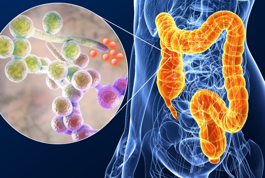
The researchers, whose report appears Oct. 14 in Cell, found in a preclinical model that pain-sensing neurons in the gut secrete a molecule called substance P, which appears to protect against gut inflammation and related tissue damage by boosting the population of beneficial microbes in the gut. The researchers also found that these pain-sensing nerves are diminished in number, with significant disruptions to their pain-signaling genes, in people who have inflammatory bowel disease (IBD).

“These findings reshape our thinking about chronic inflammatory disease, and open up a whole new approach to therapeutic intervention,” said study senior author Dr. David Artis, director of the Jill Roberts Institute for Research in Inflammatory Bowel Disease, director of the Friedman Center for Nutrition and Inflammation and the Michael Kors Professor of Immunology at Weill Cornell Medicine.

a man posing for a picture

Dr. David Artis


The study’s first author, Dr. Wen Zhang, a postdoctoral researcher in the Artis laboratory, added, “Defining a previously unknown sensory function for these specific neurons in influencing the microbiota adds a new level of understanding to host-microbiota interactions.”

IBD covers two distinct disorders, Crohn’s disease and ulcerative colitis, and is believed to affect several million people in the United States. Typically it is treated with drugs that directly target elements of the immune system. Scientists now appreciate that gut-dwelling bacteria and other microbes also help regulate gut inflammation.

As Dr. Artis’s laboratory and others have shown in recent years, the nervous system, which is “wired” into most organs, appears to be yet another powerful regulator of the immune system at the body’s barrier surfaces. In the new study, Dr. Artis and his team specifically examined pain neurons that innervate—extend their nerve endings into—the gut.

a woman smiling for a portrait

Dr. Wen Zhang


These gut-innervating pain neurons, whose cell bodies sit in the lower spine, express a surface protein called TRPV1, which serves as a receptor for pain-related signals. TRPV1 can be activated by high heat, acid, and the chili-pepper compound capsaicin, for example—and the brain translates this activation into a sense of burning pain. The researchers found that silencing these TRPV1 receptors in gut nerves, or deleting TRPV1-expressing neurons, led to much worse inflammation and tissue damage in IBD mouse models, whereas activating the receptors had a protective effect.

The investigators observed that the worsened inflammation and tissue damage in TRPV1-blocked mice were associated with changes in the relative populations of different species of gut bacteria. When this altered bacterial population was transplanted into normal mice, it caused the same worsened susceptibility to inflammation and damage. By contrast, broad-spectrum antibiotic treatment could reverse this susceptibility even in TRPV1-blocked mice. This result demonstrated that TRPV1-expressing nerves protect the gut mainly by helping to maintain a healthy gut microbe population. To continue reading, click here.

Fungal Association with Tumors May Predict Worse Outcomes

The presence of some fungal species in tumors predicts—and may even help drive—worse cancer outcomes, according to a study from Weill Cornell Medicine and Duke University researchers.

The study, which appears Sept. 29 in Cell, provides a scientific framework to develop tests that delineate specific fungal species in tumors that are relevant for prediction of cancer progression and therapy. The results also point to the possibility of using antifungal treatments to augment conventional cancer treatments in some cases.

“These findings open up a lot of exciting research directions, from the development of diagnostics and treatments to studies of the detailed biological mechanisms of fungal relationships to cancers,” said senior author Dr. Iliyan Iliev, associate professor of immunology in medicine in the Division of Gastroenterology and Hepatology and a member of the Jill Roberts Institute for Research in Inflammatory Bowel Disease at Weill Cornell Medicine.
a man posing for a picture

Dr. Iliyan Iliev. Credit: Jennifer Conrad


The first author of the study was Anders Dohlman, a doctoral student in biomedical engineering at Duke University.

The idea that viruses and bacteria can trigger or accelerate cancer development is now well established. However, little is known about the cancer-related roles of fungi—which, like bacteria and viruses, colonize the gut, lungs, skin and other barrier tissues, interact with the immune system, and sometimes cause disease.

In the new study, researchers catalogued fungal species and their associations with different cancers by analyzing The Cancer Genome Atlas, the largest well-annotated genomic database of human tumors.

The analysis revealed that the DNA of certain fungal species are relatively abundant in some tumor types. These species include, in gastrointestinal tumors, Candida tropicalis and Candida albicans, which causes thrush and yeast infections; in lung tumors, species of the fungal genus Blastomyces; and in breast tumors, species of the fungus Malassezia.

The researchers devised sophisticated computational methods to exclude fungal DNA likely to have originated from laboratory contamination, and were able to confirm, in particular, the presence of live Candida species in colorectal tumor samples.

Their analysis linked higher levels of Candida in gastrointestinal tumors to tumor gene activity promoting inflammation and reduced cell-to-cell adhesion—features associated with cancer’s late-stage spread to distant organs, known as metastasis. Higher Candida levels for such tumors were also directly associated with a greater rate of metastasis.

The findings, according to the researchers, suggest that high levels of particular fungi in tumor biopsies might someday be used as biomarkers, indicating, for example, a higher metastasis risk—which in turn could lead to the choice of more effective treatment. To continue reading, click here.

Discovery Reveals How the Immune System Tolerates Friendly Gut Bacteria

Immune cells called group 3 innate lymphoid cells (ILC3s) play an essential role in establishing tolerance to symbiotic microbes that dwell in the human gastrointestinal tract, according to a study led by researchers at Weill Cornell Medicine.

The discovery, reported Sept. 7 in Nature, illuminates an important aspect of gut health and mucosal immunity—one that may hold the key to better treatments for inflammatory bowel disease (IBD), colon cancer and other chronic disorders.

“As part of this study, we define a novel pathway that drives immune tolerance to microbiota in the gastrointestinal tract,” said senior author Dr. Gregory F. Sonnenberg, associate professor of microbiology and immunology in medicine and head of basic research in the Division of Gastroenterology & Hepatology, and a member of the Jill Roberts Institute for Research in Inflammatory Bowel Disease at Weill Cornell Medicine. “This is a fundamental advance in our understanding of mucosal immunity and may hold the key to understanding what goes wrong when the immune system begins to inappropriately attack microbiota in diseases such as IBD.”

two men posing for a photo

Drs. Mengze Lyu and Gregory Sonnenberg


Scientists have long known that trillions of bacteria, fungi, and other microbes dwell symbiotically in the intestines of mammals. The mechanism by which the immune system normally tolerates these “beneficial” gut microbes, instead of attacking them, has not been well understood. But there is evidence that this tolerance breaks down in IBD, leading to harmful flareups of gut inflammation. Thus, a detailed understanding of gut immune tolerance could enable the development of powerful new treatments for IBD—a class of diseases that include Crohn’s disease and ulcerative colitis, which affect several million individuals in the United States alone.
In the study, Dr. Sonnenberg and colleagues, including lead author Dr. Mengze Lyu, a postdoctoral researcher in the Sonnenberg lab, used single-cell sequencing and fluorescent imaging techniques to delineate immune cells in the mesenteric lymph nodes that drain the intestines of healthy mice. They focused on cells expressing a transcription factor, RORγt, which are known to drive either inflammation or tolerance in response to microbes that colonize the intestine. The dominant immune cell types in these tissues, they found, were T cells and ILC3s. The latter are a family of immune cells that represent an innate counterpart of T cells, and work as a first line of defense in mucosal tissues such as the intestines and lungs.

In close collaboration with researchers at the University of Birmingham, UK, the scientists observed that in lymph node regions called interfollicular zones, ILC3s are in close association with a specific type of T cell, called RORγt+ regulatory T cells (Tregs), which are adapted to dial down inflammation and immune activity to promote tolerance in the gut. To continue reading, please click here.

Scientists Identify a Key Molecular Protector of Gut Health

A protein called Zbtb46, expressed by specialized immune cells, has a major role in protecting the gastrointestinal tract from excessive inflammation, according to a study from researchers at Weill Cornell Medicine.

The finding, which appears July 13 in Nature, is a significant advance in the understanding of how the gut maintains health and regulates inflammation, which could lead to better strategies for treating diseases like inflammatory bowel disease (IBD).

“We’ve known that there are related families of immune cells in the gut that can either protect from inflammation or at other times be major drivers of inflammation,” said senior author Dr. Gregory Sonnenberg, associate professor of microbiology and immunology in medicine in the Division of Gastroenterology & Hepatology, and a member of the Jill Roberts Institute for Research in Inflammatory Bowel Disease at Weill Cornell Medicine. “This new finding helps us understand how these cells are regulated to optimally promote intestinal health and prevent inflammation.”

IBD, which includes Crohn’s disease and ulcerative colitis, affects several million people in the United States. These chronic inflammatory disorders target the gut, can be seriously debilitating, and treatments may not work well for some patients—mainly because scientists don’t have a complete picture of what is driving these diseases and how the sophisticated immune cell networks in the gut support tissue health.

Dr. Sonnenberg’s laboratory has been advancing the science of gut immunity with studies of recently identified immune cells called ILC3s. These “innate lymphoid cells” are related to T cells and B cells, and clearly have important roles in protecting the gut and other organs from excess inflammation. However, in the context of IBD or colorectal cancer they become altered. In general, scientists have wanted to know more about how these cells work.

a man and woman smiling for a portrait

Drs. Gregory Sonnenberg and Wenqing Zhou. Provided by Sonnenberg lab


To this end, Dr. Sonnenberg and his team, including first author Dr. Wenqing Zhou, a postdoctoral researcher in the Sonnenberg Laboratory, set out to make a detailed catalog of ILC3s and other related immune cells residing in the large intestine of mice, using relatively new single-cell sequencing techniques.

A surprise finding was that a subset of ILC3s express Zbtb46, an anti-inflammatory protein that prior studies had suggested is produced only in dendritic cells, a very different type of immune cell. The researchers showed in experiments with mice that ILC3s expressing Zbtb46 have a strong ability to restrain inflammation following gut infection. When they blocked Zbtb46 expression in ILC3s, gut infection led to signs of severe inflammation, including a rise in the numbers of other gut immune cells promoting inflammation. To read further, please click here

Toxin-producing Yeast Strains in Gut Fuel IBD

Individual Candida albicans yeast strains in the human gut are as different from each other as the humans that carry them, and some C. albicans strains may damage the gut of patients with inflammatory bowel disease (IBD), according to a new study from researchers at Weill Cornell Medicine. The findings suggest a possible way to tailor treatments to individual patients in the future.

The researchers, who report their findings March 16 in Nature, used an array of techniques to study strains, or genetic variants, of Candida from the colons of people with or without ulcerative colitis, a chronic, relapsing and remitting inflammatory disorder of the colon and rectum and one of the main forms of IBD. They found that certain strains, which they call “high-damaging,” produce a potent toxin called candidalysin that damages immune cells.

a man posing for a picture

Dr. Iliyan Iliev


“Such strains retained their “high-damaging” properties when they were removed from the patient’s gut and triggered pro-inflammatory immunity when colonized in mice, replicating certain disease hallmarks,” said senior author Dr. Iliyan Iliev, an associate professor of immunology in medicine in the Division of Gastroenterology and Hepatology and a scientist in the Jill Roberts Institute for Research in Inflammatory Bowel Disease at Weill Cornell Medicine.

IBD affects approximately 3.1 million people in the United States and can greatly impair patients’ quality of life. Such patients rely on a handful of available therapies, but treatments may not always be effective. The new study has suggested one reason steroids, a commonly used treatment, may not work; treating mice with the drug to suppress intestinal inflammation failed in the presence of “high-damaging” C. albicans strains.

“Our findings suggest that C. albicans strains do not cause spontaneous intestinal inflammation in a host with intact immunity,” Dr. Iliev said. “But they do expand in the intestines when inflammation is present and can be a factor that influences response to therapy in our models and perhaps in patients.”

Most studies of the human microbiome in healthy individuals and those with IBD have focused on bacteria and viruses, but recent research by Dr. Iliev and others has illuminated the contributions of fungi to the effects of microbes on humans and mice. They have found that intestinal fungi play an important role in regulating immunity at surfaces exposed to the outside, such as the intestines and lungs, due to their potent immune-stimulating characteristics. While the collective community of fungi in the body, known as the mycobiota, has been linked to several diseases, including IBD, researchers previously had not understood the mechanisms by which the mycobiota contribute to inflammation in the gut. To continue reading article, click here.

A New and Versatile Genetic Manipulation Pipeline for Studying Nonmodel Gut Bacteria

Scientists at the Jill Roberts Institute for Research in Inflammatory Bowel Disease at Weill Cornell Medicine have developed a pipeline that enables genetic manipulation of nonmodel gut bacteria. The pipeline will allow scientists to study the biological roles of these bacteria, which are increasingly recognized as key factors in health and disease, at the single-gene level.

Scientists have developed advanced genetic tools for some model gut bacteria, such as E. coli, but have lacked the necessary tools for a large group of gut bacteria called Firmicutes/Clostridia that are dominant in a healthy human gut. In the study, published Jan. 19 in Cell, the researchers have developed gene-modification techniques for multiple nonmodel gut bacteria, from more than five different phyla including Firmicutes/Clostridia.

The researchers demonstrated the potential of their new genetic tools by using them to study the role of a key gut bacterial gene in regulating colon inflammation.

"Gut bacteria play a major role in the onset and development of human diseases like inflammatory bowel disease and cancers," said Dr. Chun-Jun Guo, assistant professor of immunology in medicine in the Department of Gastroenterology and Hepatology and a scientist in the Jill Roberts Institute for Research in Inflammatory Bowel Disease at Weill Cornell Medicine. “We were able not only to develop genetic manipulation tools for previously 'intractable' gut bacteria, but also to discover an interesting role for a microbial gene in the host using these tools.”

Gut bacteria have generally evolved symbiotic relationships, also known as commensal, with their animal hosts, including humans. Although scientists over the last two decades have been increasingly aware of gut microbes' importance in immune function and overall health—and in promoting disease when they are disrupted, for example by antibiotic use or poor diets—the development of genetic tools for manipulating these microbes has not kept pace. To read further, click here.

Two Weill Cornell Medicine Faculty Members Inducted into ASCI

Two Weill Cornell Medicine physician-scientists, Dr. Randy Longman and Dr. Robert Schwartz, have been elected as members of the American Society for Clinical Investigation.

The American Society for Clinical Investigation (ASCI) is one of the nation’s oldest nonprofit medical honor societies and focuses on the unique role of physician-scientists in research, clinical care and medical education. It is comprised of more than 3,000 physician-scientists serving in the upper ranks of academic medicine and industry. Members are leaders in their fields in translating innovative laboratory findings into clinical advancements. Drs. Longman and Schwartz join 93 other new members elected this year and will be officially inducted at the organization’s annual meeting in April.

a man in a suit posing for a picture

Dr. Randy Longman


Dr. Randy Longman is an associate professor of medicine in the Division of Gastroenterology and Hepatology at Weill Cornell Medicine and director of the Jill Roberts Center for Inflammatory Bowel Disease at Weill Cornell Medicine and NewYork-Presbyterian/Weill Cornell Medical Center. His research focuses on interactions between gut microbes and the immune system in inflammatory bowel disease (IBD). His work has identified specific strains of bacteria that are involved and how they relay signals to the immune system to activate inflammation in patients with Crohn’s disease and ulcerative colitis, the most common forms of IBD.

Dr. Longman is currently leading a clinical trial evaluating whether dietary fiber supplementation may improve the efficacy of fecal microbiota transplantation (FMT) in patients with moderate ulcerative colitis. Supported by the National Institute of Diabetes and Digestive and Kidney Diseases, the trial is based on his earlier work that identified specific microbes associated with clinical responses to FMT. Dr. Longman’s research is funded by several organizations, including the National Institutes of Health and the Crohn’s and Colitis Foundation.

“It’s very meaningful to be elected as a member of the ASCI, knowing my peers recognize my work has contributed toward moving the needle forward,” said Dr. Longman, who is also a member of the Jill Roberts Institute for Research in Inflammatory Bowel Disease. “As a physician-scientist, the most important thing for me is that my research discoveries may translate into clinical treatments that help patients.” To read further, click here.

Preclinical Study Finds Gut Fungi Influence Neuroimmunity and Behavior

A specific group of fungi residing in the intestines can protect against intestinal injury and influence social behavior, according to new preclinical research by scientists at Weill Cornell Medicine. The findings extend a growing body of work identifying a "gut-immunity-brain axis," a signaling system that may have a wide range of effects on physiology in both health and disease, influenced not only by the body's own cells but also the resident microbes.

The study, published Feb. 16 in Cell, reveals a novel set of molecular signals connecting fungi in the gut to their host’s cells throughout the body, including immune cells and neurons.

“We have made a direct link between a major immune pathway induced by fungi in the lining of the intestine and signals in the nervous system that impact animal behavior,” said senior author Dr. Iliyan Iliev, associate professor of immunology in medicine in the Division of Gastroenterology and Hepatology and a member of the Jill Roberts Institute for Research in Inflammatory Bowel Disease at Weill Cornell Medicine.

Drs. Dilek ColakMelanie Johncilla and Megan Allen from Weill Cornell Medicine and Dr. Rhonda K. Yantiss from Weill Cornell Medicine and NewYork-Presbyterian also contributed to this study.

The lining of the intestine must balance conflicting needs, absorbing water and nutrients from food while acting as a barrier to prevent the vast population of microbes in the gut from invading the bloodstream. Examining this system in a mouse model, the scientists mapped the locations of different fungi within the intestine and found that a unique consortium of fungi tends to accumulate at specific sites near the gut epithelium, or lining, suggesting that these species have colonized the gut and interact closely with the nearby epithelial cells.

image of two people in front of flow cytometry

Dr. Irina Leonardi (left) and Dr. Iliyan Iliev. Photo credit: Jennifer Conrad.


Mice carrying some of these fungi enjoyed better protection against events that can disrupt the intestinal barrier, such as intestinal injury and bacterial infection. "There was fortification of those barrier functions when we added that specific fungal community to mice," Dr. Iliev said.

Improving intestinal barrier integrity wasn't the only effect of the fungi. In separate experiments, the team found that mice carrying the fungal community in their gut displayed more social behavior than animals without these fungi. To read further, click here.

Key Growth Factor Protects Gut from Inflammatory Bowel Disease 

microscopic gut cells

Image demonstrating TNF-driven intestinal inflammation, where the gut-lining epithelial cells (marked by Epcam in green) exhibit substantial cell death (marked by cleaved caspase 3 in red). Image courtesy of Dr. Lei Zhou.

A growth factor protein produced by rare immune cells in the intestine can protect against the effects of inflammatory bowel disease (IBD), according to a new discovery from Weill Cornell Medicine researchers.

In their study, published Jan. 31 in Nature Immunology, the researchers found that the growth factor, HB-EGF, is produced in response to gut inflammation by a set of immune-regulating cells called ILC3s. These immune cells reside in many organs including the intestines, though their numbers are known to be depleted in the inflamed intestines of IBD patients.

The researchers showed in experiments in mice that this growth factor can powerfully counter the harmful effects of a key driver of intestinal inflammation called TNF. In doing so, ILC3s protect gut-lining cells when they would otherwise die and cause a breach in the intestinal barrier.

a man in a suit posing for a picture

Dr. Gregory Sonnenberg


“We’ve discovered a new cellular pathway that is essential to protect against gut inflammation. This discovery could lead to a better understanding of IBD pathogenesis and new strategies to treat this disease” said study senior author Dr. Gregory Sonnenberg, an associate professor of microbiology and immunology in medicine in the Division of Gastroenterology & Hepatology and a scientist in the Jill Roberts Institute for Research in Inflammatory Bowel Disease at Weill Cornell Medicine.

IBD, a disease category including ulcerative colitis and Crohn’s disease, features chronic gut inflammation and many potential follow-on effects including arthritis and colorectal cancer. The condition appears to be quite common in the United States; a survey-based study by researchers at the Centers for Disease Control and Prevention in 2015 suggested that more than 1 percent of the U.S. population—more than three million people—were living with IBD. Current treatments help some but not all patients.

Dr. Sonnenberg and his laboratory have found in recent studies that ILC3s play a key role in protecting the gut from harmful inflammation and are depleted in human patients who have IBD or colon cancer. In the new study, the team sought a more precise understanding of how ILC3s fight against IBD’s inflammatory effects.

The researchers in an initial set of experiments recreated an IBD-like condition in mice using high doses of an inflammatory immune protein called TNF, a major driver of inflammation in IBD and the target of some IBD therapies. They found in these experiments that ILC3s strongly protect the gut linings of the mice from TNF-induced inflammatory damage—mice lacking ILC3s suffered significantly worse damage.

Prior studies have suggested that ILC3s help protect the gut at least in part by producing an immune protein called IL-22, which promotes gut barrier function. However, the mouse experiments in the new study indicated that ILC3s’ gut-protecting effect against TNF works independently of IL-22.

Using a relatively advanced technique called single-cell RNA sequencing, the researchers eventually zeroed in on the mechanism of ILC3s’ protective effect: the growth factor protein HB-EGF, which they showed could specifically keep gut-lining cells alive in the presence of excessive TNF.

The team found that ILC3s are the dominant producers of HB-EGF in the gut. They were able to identify the cascade of signaling factors that occurs downstream of TNF and causes ILC3 to switch on HB-EGF production—and they observed the same cascade in human ILC3s, indicating that these findings are not just specific to mice. The researchers also confirmed from analyses of IBD-patient gut tissue that HB-EGF-producing ILC3s are reduced in areas of gut inflammation.

The findings reveal a key mechanism that the gut normally uses to protect itself from harmful inflammation and suggest that the loss of ILC3s is at least one reason this mechanism fails in IBD.

“Identifying the significance of this pathway is a good first step, and we’re now thinking about how we might manipulate this pathway to benefit IBD patients,” Dr. Sonnenberg said.

The loss of ILC3s in the IBD gut poses a challenge to the development of therapeutic solutions that depend on ILC3s, he noted. Moreover, though the growth factor HB-EGF on its own could be therapeutic, even if ILC3s are depleted, HB-EGF has been linked to the faster growth of a variety of cancers.

a man posing for a picture

Dr. Lei Zhou


“Our ongoing research is interrogating the role of this ILC3 and HB-EGF pathway in the development of chronic-inflammation-related colon cancer,” said study first author Dr. Lei Zhou, a postdoctoral associate in the Sonnenberg laboratory. “It will be important to delineate the exact cellular and molecular mechanisms by which this novel pathway coordinates intestinal health, inflammation and cancer before moving forward with manipulating it as a therapeutic strategy.”

The Sonnenberg Laboratory is supported by the National Institutes of Health (R01AI143842, R01AI123368, R01AI145989, R01AI162936, R21CA249284 and U01AI095608), the NIAID Mucosal Immunology Studies Team (MIST), the Crohn’s and Colitis Foundation, the Searle Scholars Program, the American Asthma Foundation Scholar Award, Pilot Project Funding from the Center for Advanced Digestive Care (CADC), an Investigators in the Pathogenesis of Infectious Disease Award from the Burroughs Wellcome Fund, a Wade F.B. Thompson/Cancer Research Institute (CRI) CLIP Investigator grant, the Meyer Cancer Center Collaborative Research Initiative, the Dalton Family Foundation, Linda and Glenn Greenberg, and the Roberts Institute for Research in IBD. Gregory F. Sonnenberg is a CRI Lloyd J. Old STAR. Lei Zhou is supported by a fellowship from the Crohn’s and Colitis Foundation (608975).

Weill Cornell Medicine Friedman Center for Nutrition & Inflammation 413 E. 69th Street, Seventh Floor New York, NY 10021